Generalized Additive Modeling Helps Identify Unique Brain Regions Affected By Major Depressive Disorder
Worldwide, hundreds of millions of people spanning adulthood are afflicted with Major Depressive Disorder (MDD), and diagnosed cases have increased in recent years. In the past, the search for valuable fluid biomarkers for use in clinical diagnosis has been limited and is still in the early discovery stages. Structural brain changes have been suggested to impact MDD, but regional detail is largely unexplored. Dr Tosun-Turgut and colleagues used neuroimaging data to analyze changes in brain structure in regions known to be associated with MDD, and correlated these regional changes with age.

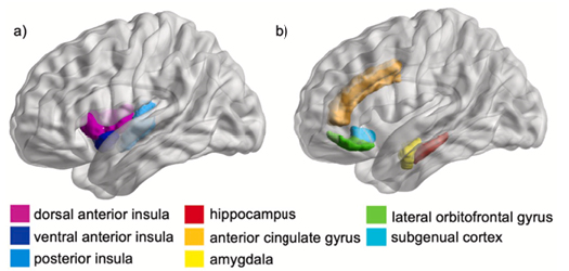
This study compared T1-weighted magnetic resonance (MR) images from unmedicated MDD patients with images from healthy control participants (CTLs) from 18-85 years old. ComBat-GAM was used to harmonize imaging data and then looked for changes in specific regions of interest of the insula (above, left) as compared to brain regions implicated in MDD (above, right).
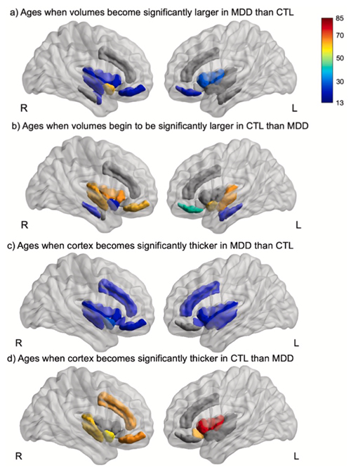
Modeling revealed differences in gray matter (GM) volume and cortical thickness measures as young as 18 yrs old. They found that as all subjects age, the brain has smaller GM volume and thinner cortical tissue. In the lateral orbital gyrus and all insular subregions, MDD patients had greater GM volume and thicker cortices from ~18-35 yrs, but smaller GM volume and thinner cortices during and after middle age.

This work has shown that differences in brain morphometry (size and shape) exist between MDD patients and non-MDD patients and change with age. These changes are found in regions associated with MDD and may help with future diagnostics.
Notably, the MR images in this study were from four different cohorts; harmonization of data allowed researchers to enrich their dataset. This method will impact all types of neuroimaging studies – longitudinally and cross-sectionally - by normalizing images that may have been acquired at different times, on different MR imaging machines, and at different sites throughout the years.
This paper was published in Neuroimage: Clinical. Additional authors include Adam Lang (NCIRE), Charles Taylor (UCSD), R. Scott Mackin (NCIRE/UCSF), Dieter Meyerhoff (NCIRE/UCSF), Susanne Mueller (NCIRE/UCSF), and Irina Strigo (UCSF/SFVAHCC).

